Mohit Adhikari, Belgium
Bio-Imaging Lab, University of Antwerp Biomedical SciencesAuthor Of 3 Presentations
LIVE DISCUSSION
SPATIOTEMPORAL ALTERATIONS OF RESTING-STATE QUASI-PERIODIC PATTERNS IN 4-MONTH OLD TGF344-AD RATS
Abstract
Aims
Alzheimer’s Disease (AD) is a severe neurodegenerative disorder that leads to brain network dysfunction and cognitive decline. Changes in functional networks at symptomatic stages of AD can be captured using resting state (RS) fMRI in patients and animal models. Here, we used a rat model manifesting the full-spectrum of human AD-pathology to identify if spatiotemporal network alterations are present at an early, pre-symptomatic stage.
Methods
RS fMRI data were collected from four-month-old TgF344-AD rats (N=15) and wildtype (WT) (N=11) littermates. Acquired images were realigned, normalized to a 3D- template, masked, smoothed, filtered and the global signal was regressed out. Recurrent patterns of brain activity (3.6 seconds long) were extracted using quasi-periodic pattern (QPP) analysis starting from 200 different seed patterns. Then, the 200 patterns of each group were clustered based on temporal and spatial similarity to identify the representative QPPs based on occurrence rates.
Results
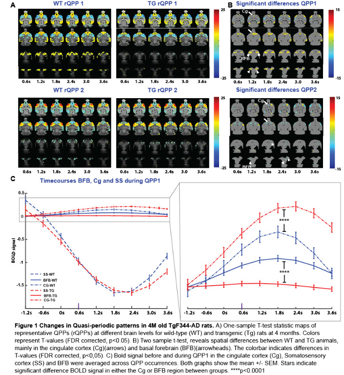
Voxel-wise activations of matched QPPs across groups (Fig 1A)were compared using a two-sample t-test, FDR corrected for multiple comparisons. Significant QPP differences between groups were primarily found in the basal forebrain (BFB), and cingulate cortex (Cg) (Fig 1B). QPP time-course analysis in these regions-of-interest and somatosensory cortex (SS), demonstrated a concomitant reduction of BFB and an increase of Cg activity in the Tg-rats compared to the WT controls, while SS activity profiles remained unchanged (Fig 1C).
Conclusions
In summary, our results highlight the important role of the BFB in regulating whole-brain networks and indicate a potential signature to identify early onset changes at the network level.
RESTING-STATE CO-ACTIVATION PATTERNS AS ACCURATE PREDICTORS OF LATE-STAGE ALZHEIMER’S DISEASE IN MOUSE MODELS.
Abstract
Aims
Alzheimer’s disease (AD), a neurodegenerative disorder marked by accumulation of extracellular amyloid-beta (Aβ) plaques leads to progressive loss of memory and cognitive function. Resting-state fMRI (RS-fMRI) studies have provided links between these observations in terms of disruption of default mode and task-positive resting-state networks (RSNs). The objective of our study was to investigate dynamic fluctuations in RS-fMRI signals in a mouse model (TG2576) of amyloidosis at a late stage (18-months) using sets of resting-state co-activation patterns (CAPs) & to assess their predictive ability.
Methods
CAPs are transient patterns of co-(de)activation that form the basis of RSNs. Here, we followed the approach by Gutierrez-Barragan et al. (2019) that divided all time frames within a scan into spatially similar clusters to extract CAPs from RS-fMRI in 10 TG2576 female mice and 8 wild-type (WT) littermates. Subsequently, we matched the CAPs from two groups using the Hungarian method & compared their temporal (duration, occurrence rate) and the spatial (lateralization of significantly activated voxels) properties. Finally, we tested the predictive ability of CAPs by using their spatial & temporal components as features for training a classifier in order to distinguish the transgenic mice from the WT.
Results
We found robust differences in the spatial component of matched CAPs. We found that while their duration & occurrence rates classified significantly better than the chance-level, BOLD intensities of significantly activated voxels of CAPs turned out to be a significantly better predictive feature achieving a 90% accuracy.
Conclusions
We conclude that RS-CAPs can serve as diagnostic and, potentially, prognostic biomarkers of Alzheimer's disease.
Presenter of 2 Presentations
RESTING-STATE CO-ACTIVATION PATTERNS AS ACCURATE PREDICTORS OF LATE-STAGE ALZHEIMER’S DISEASE IN MOUSE MODELS.
Abstract
Aims
Alzheimer’s disease (AD), a neurodegenerative disorder marked by accumulation of extracellular amyloid-beta (Aβ) plaques leads to progressive loss of memory and cognitive function. Resting-state fMRI (RS-fMRI) studies have provided links between these observations in terms of disruption of default mode and task-positive resting-state networks (RSNs). The objective of our study was to investigate dynamic fluctuations in RS-fMRI signals in a mouse model (TG2576) of amyloidosis at a late stage (18-months) using sets of resting-state co-activation patterns (CAPs) & to assess their predictive ability.
Methods
CAPs are transient patterns of co-(de)activation that form the basis of RSNs. Here, we followed the approach by Gutierrez-Barragan et al. (2019) that divided all time frames within a scan into spatially similar clusters to extract CAPs from RS-fMRI in 10 TG2576 female mice and 8 wild-type (WT) littermates. Subsequently, we matched the CAPs from two groups using the Hungarian method & compared their temporal (duration, occurrence rate) and the spatial (lateralization of significantly activated voxels) properties. Finally, we tested the predictive ability of CAPs by using their spatial & temporal components as features for training a classifier in order to distinguish the transgenic mice from the WT.
Results
We found robust differences in the spatial component of matched CAPs. We found that while their duration & occurrence rates classified significantly better than the chance-level, BOLD intensities of significantly activated voxels of CAPs turned out to be a significantly better predictive feature achieving a 90% accuracy.
Conclusions
We conclude that RS-CAPs can serve as diagnostic and, potentially, prognostic biomarkers of Alzheimer's disease.


