Laura Wisse, Sweden
Lund University Diagnostic RadiologyPresenter of 2 Presentations
LIVE DISCUSSION
HIGH-RESOLUTION POSTMORTEM MRI REVEALS PATHOLOGY-SPECIFIC NEURODEGENERATION PATTERNS IN THE MEDIAL TEMPORAL LOBE
Abstract
Aims
The medial temporal lobe (MTL) is a hotspot for neurodegenerative pathologies and therefore an important region to study polypathology. We investigate the association of different neurodegenerative pathologies and the thickness of different granular MTL subregions measured on high-resolution postmortem MRI.
Methods
Tau, TDP-43, β-amyloid and α-synuclein pathology were rated (0-absent – 3-frequent) in the hippocampus and entorhinal cortex (ERC) of 58 individuals with and without neurodegenerative diseases (mean age 74.7 years, 39.7% female). Thickness measurements using a semi-automated approach were obtained from 0.2x0.2x0.2 mm3 post-mortem MRI scans of excised MTL specimens from the contralateral hemisphere in the ERC, Brodmann Area (BA) 35 and 36, parahippocampal cortex (PHC), subiculum (SUB), cornu ammonis (CA)1 and the stratum radiatum lacunosum moleculare (SRLM). Spearman’s rank correlations were performed, correcting for age, sex and hemisphere, including all four proteinopathies in the model.
Results
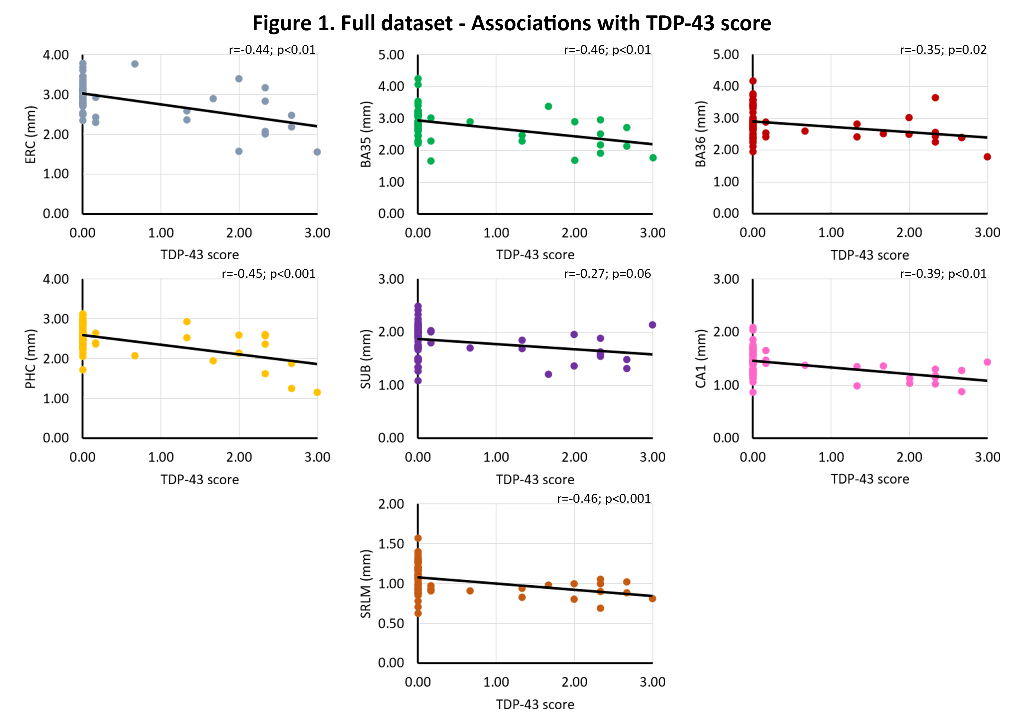
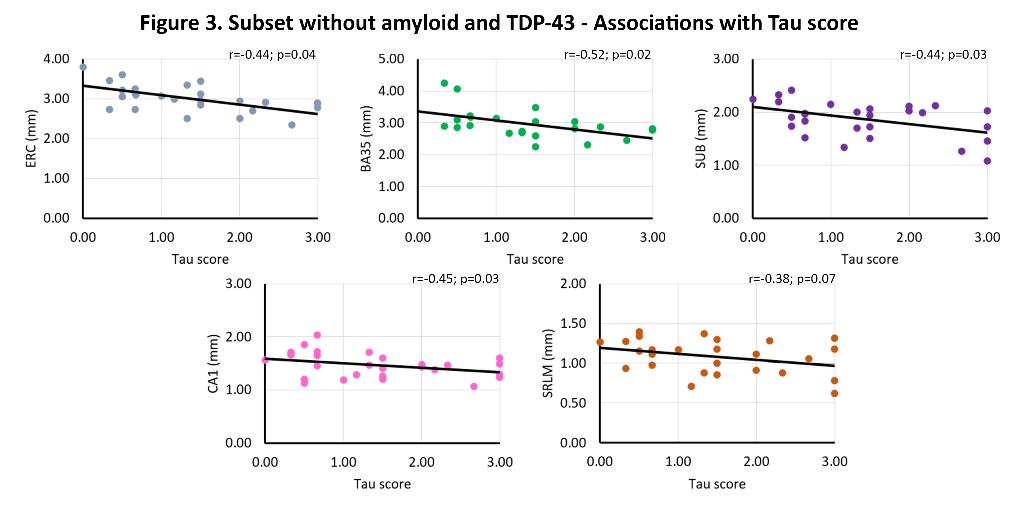
We find significant associations of 1) TDP-43 with all subregions (r=-0.27 – r=-0.46, trend for subiculum), and 2) tau with ERC (r=-0. 26, trend), BA35 (r=-0.31) and SRLM (r=-0.33). In β-amyloid and TDP-43 negative cases, we find strong significant associations of tau with ERC (r=-0.40, trend), BA35 (r=-0.55), subiculum (r=-0.42), CA1 (r=-0.47) and SRLM (r=-0.38, trend). See figures 1-3.


Conclusions
This unique dataset showed widespread atrophy in relation to TDP-43 pathology and atrophy in early Braak regions and tau pathology. Moreover, the strong association of tau with thickness in early Braak regions in the absence of β-amyloid and TDP-43 is indicative for a role of Primary Age Related Tauopathy in neurodegeneration.


