Welcome to the AD/PD™ 2021 Interactive Program
The congress will officially run on Central European Time (CET) - Barcelona Time
To convert the congress times to your local time Click Here
Icons Legend: ![]()
 - Live Session |
  - On Demand Session |
  - On Demand with Live Q&A
The viewing of sessions, cannot be accessed from this conference calendar. All sessions are accessible via the Main Lobby.
FOLLOWING THE LIVE DISCUSSION, THE RECORDING WILL BE AVAILABLE IN THE ON-DEMAND SECTION OF THE AUDITORIUM.
MRI AS A TOOL TO STUDY MICROSTRUCTURAL CHANGES IN AGING AND DEMENTIA
Abstract
Abstract Body
Various MRI techniques including diffusion tensor imaging (DTI) and magnetization transfer imaging as well as iron mapping allow to detect changes in tissue microstructure in Alzheimer´s disease. The contribution of these techniques as diagnostic and prognostic tools are widely undetermined. This presentation will display new data on free water imaging, Magnetization Transfer Imaging and Iron Mapping.
Recent advances in diffusion MRI modeling enable more detailed insight into DTI alterations. Of specific interest is the free water (FW) diffusion MRI model. In a large cohort of community-dwelling individuals we show that the FW compartment correlates more closely to cognitive impairment than traditional DTI metrics such as mean diffusivity and fractional aniosptropy. As to whether alterations of the free water compartment can predict conversion of patients to dementia is yet unclear.
The second section of the talk focuses on magnetization transfer imaging and demonstrates that reductions of the magnetization transfer ratio in AD signature areas and in white matter regions of the brain contribute to cognitive impairment of AD patients, independently of brain atrophy.
Finally, longitudinal data on MR-detected iron accumulation in AD will be shown. The finding that iron in the temporal cortex predicts cognitive decline beyond what is determined by development of brain atrophy is novel.
In conclusion, the presentation highlights the potential of various MRI techniques to determine microstructural tissue alterations in the brain of elderly people without and with dementia and indicates that such changes provide clinically relevant information beyond what can be expected from structural MRI.
HIPPOCAMPAL MEAN DIFFUSIVITY IS SENSITIVE TO ALZHEIMER PATHOLOGY IN HIPPOCAMPAL SUBREGIONS.
Abstract
Aims
Hippocampal atrophy is the most established structural imaging biomarker for Alzheimer’s disease (AD). Nevertheless, some argue that hippocampal diffusion is a more sensitive imaging marker. We addressed the pathological sensitivity of hippocampal MRI volume and diffusivity vis-à-vis, and determined the relative contribution of AD related pathological proteins in hippocampal sub-regions.
Methods
Ten clinically amnestic and pathological confirmed AD, and ten age- and gender matched control decedents underwent post-mortem in-situ 3T MRI; 3DT1 for FSL FIRST hippocampus segmentation, and DTI to obtain mean diffusivity (MD). At autopsy, hippocampal tissue was dissected and processed for Aβ and p-tau immunohistochemistry, and area % immunoreactivity in hippocampal sub-regions was calculated. MRI-pathology associations were assessed with linear mixed models and with partial correlations, corrected for age, gender and post-mortem delay.
Results
Compared to controls, AD donors showed significantly increased hippocampal MD (p<0.021), but not decreased volume (p=0.195) which was more sensitive to within-group variation. Hippocampal MD associated with Braak stage (r=0.54, p=0.033) and Thal phase (r=0.51, p=0.045), volume was not significant due to within-group variation in disease duration. In whole-group analysis, p-tau contributed to hippocampal volume (p<0.001), while both Aβ (p=0.014) and p-tau (p<0.001) contributed to hippocampal MD. Regionally, hippocampal volume was associated with Aβ in CA4, CA1 and subiculum (0.03<p<0.05), and with p-tau in DG, CA4, CA3 and CA2 (0.001<p<0.05). Hippocampal MD associated with Aβ and p-tau in all subregions (0.001<p<0.05).
Conclusions
Hippocampal MD showed to be a more robust measure with greater sensitivity to (sub-regional) AD related pathological load than hippocampal volume.
MR MORPHOMETRIC MEASURES MAY PREDICT THE SEVERITY OF NEUROPSYCHIATRIC SYMPTOMS IN THE ELDERLY
Abstract
Aims
Neuropsychiatric symptoms are frequent in dementia. Occurrence of neuropychiatric symptoms and their severity might be associated with certain morphological changes. MRI measures might be used to demonstrate the risk of neuropsychiatric symptoms.
Methods
Neuropsychiatric features as assessed with Neuropsychiatric Inventory, Modified Mini Mental State (3MS) and MRI data were utilized. We evaluated classification performance of volumetric data in patients with no/mild vs. moderate/severe neuropsychiatric features. We applied both random forest (RF) using hold-out validation approach and SVM. Then we examined the predictive performance of the model using performance measures. Lastly, using residuals approach we determined the variables with the highest predictive value. The study was funded by TUBITAK 214S048, Psychiatry Associaton of Turkey and Hacettepe University.
Results
Data of a total of 175 patients over the age of 55 were analyzed. PCA revealed volume, thickness and ventricle sizes as three components. When these were combined with the clinical data including age, gender, education and 3MS scores in order to predict neuropsychiatric symptom severity, performance of RF was proved to be higher than SVM with an accuracy of 0.93 (0.84-0.98). 3MS scores, volumes, thickness, age and education predicted the symptom severity. Potentially predictive MR parameters as determined via RF were volumes of left parahippocampus, amygdala, insula and entorhinal cortex as well as thickness of left and right parahippocampus, left and right medial orbitofrontal cortices, right entorhinal cortex and left temporal pole.
Conclusions
Certain MRI measures may be useful in predicting the development of neuropsychiatric symptoms.
HIGH-RESOLUTION POSTMORTEM MRI REVEALS PATHOLOGY-SPECIFIC NEURODEGENERATION PATTERNS IN THE MEDIAL TEMPORAL LOBE
Abstract
Aims
The medial temporal lobe (MTL) is a hotspot for neurodegenerative pathologies and therefore an important region to study polypathology. We investigate the association of different neurodegenerative pathologies and the thickness of different granular MTL subregions measured on high-resolution postmortem MRI.
Methods
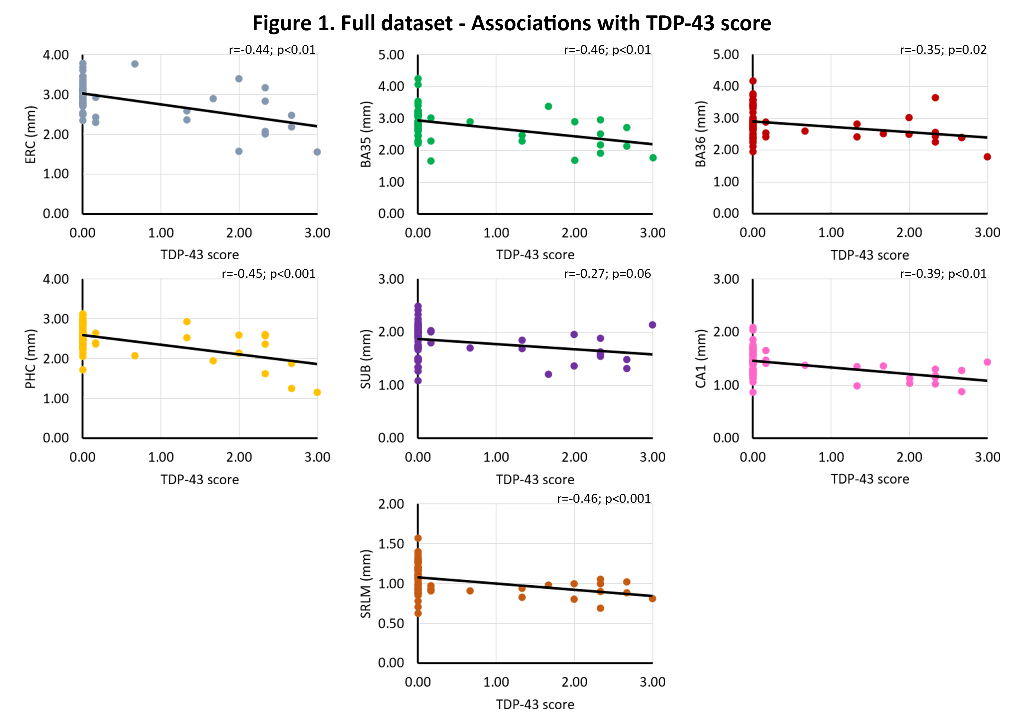
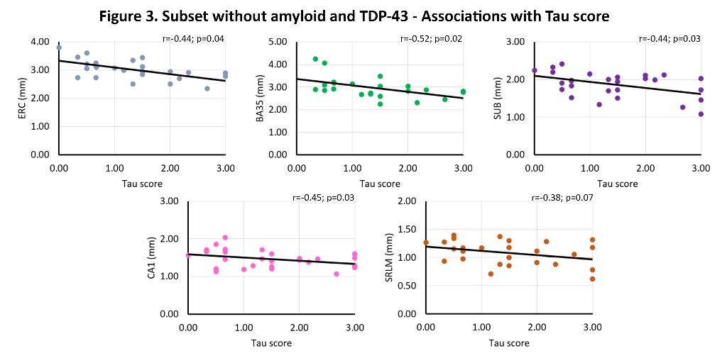
Tau, TDP-43, β-amyloid and α-synuclein pathology were rated (0-absent – 3-frequent) in the hippocampus and entorhinal cortex (ERC) of 58 individuals with and without neurodegenerative diseases (mean age 74.7 years, 39.7% female). Thickness measurements using a semi-automated approach were obtained from 0.2x0.2x0.2 mm3 post-mortem MRI scans of excised MTL specimens from the contralateral hemisphere in the ERC, Brodmann Area (BA) 35 and 36, parahippocampal cortex (PHC), subiculum (SUB), cornu ammonis (CA)1 and the stratum radiatum lacunosum moleculare (SRLM). Spearman’s rank correlations were performed, correcting for age, sex and hemisphere, including all four proteinopathies in the model.
Results
We find significant associations of 1) TDP-43 with all subregions (r=-0.27 – r=-0.46, trend for subiculum), and 2) tau with ERC (r=-0. 26, trend), BA35 (r=-0.31) and SRLM (r=-0.33). In β-amyloid and TDP-43 negative cases, we find strong significant associations of tau with ERC (r=-0.40, trend), BA35 (r=-0.55), subiculum (r=-0.42), CA1 (r=-0.47) and SRLM (r=-0.38, trend). See figures 1-3.


Conclusions
This unique dataset showed widespread atrophy in relation to TDP-43 pathology and atrophy in early Braak regions and tau pathology. Moreover, the strong association of tau with thickness in early Braak regions in the absence of β-amyloid and TDP-43 is indicative for a role of Primary Age Related Tauopathy in neurodegeneration.
FIRST-LEVEL ASSESSMENT OF NEURODEGENERATION USING DEEP-LEARNING BASED BRAIN CT SEGMENTATIONS AND BLOOD-DERIVED MEASURES OF NEUROFILAMENT LIGHT
Abstract
Aims
Brain computed tomography (CT) and blood sampling are two routinely performed examinations in the clinical assessment of neurodegenerative diseases. We aim to evaluate the diagnostic performance of automated CT-based volumetry, derived with a novel deep-learning approach, and plasma-based neurofilament light chain (NfL).
Methods
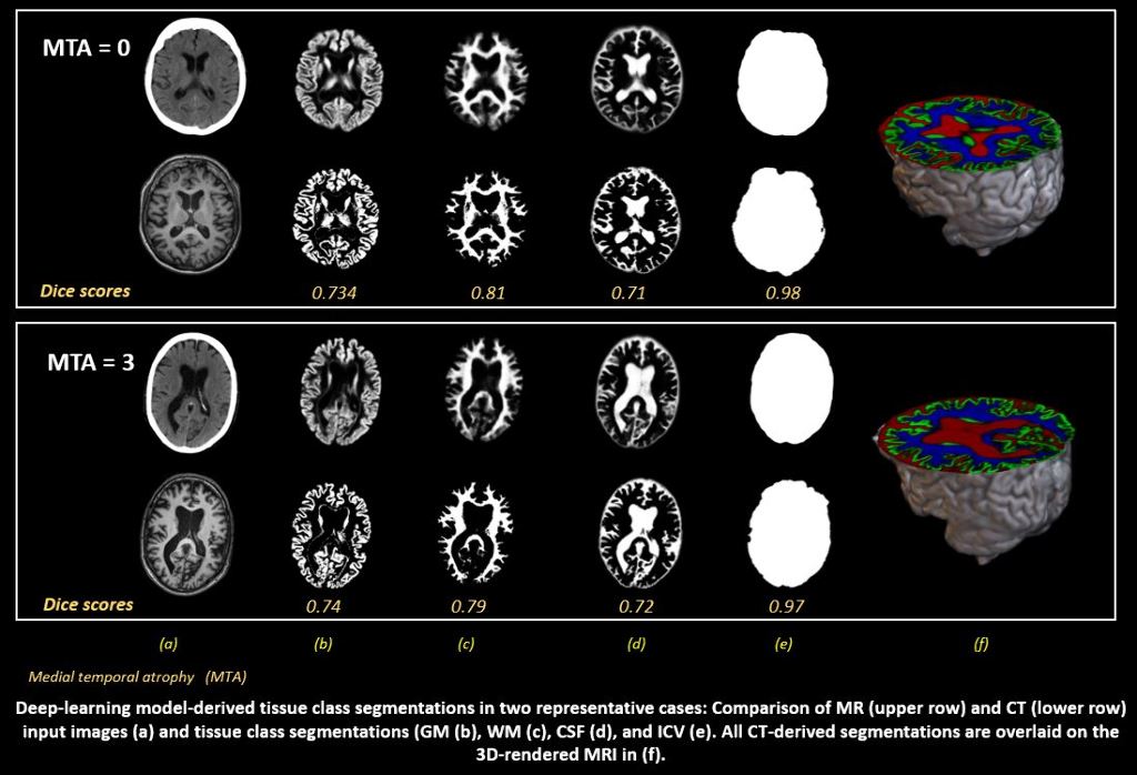
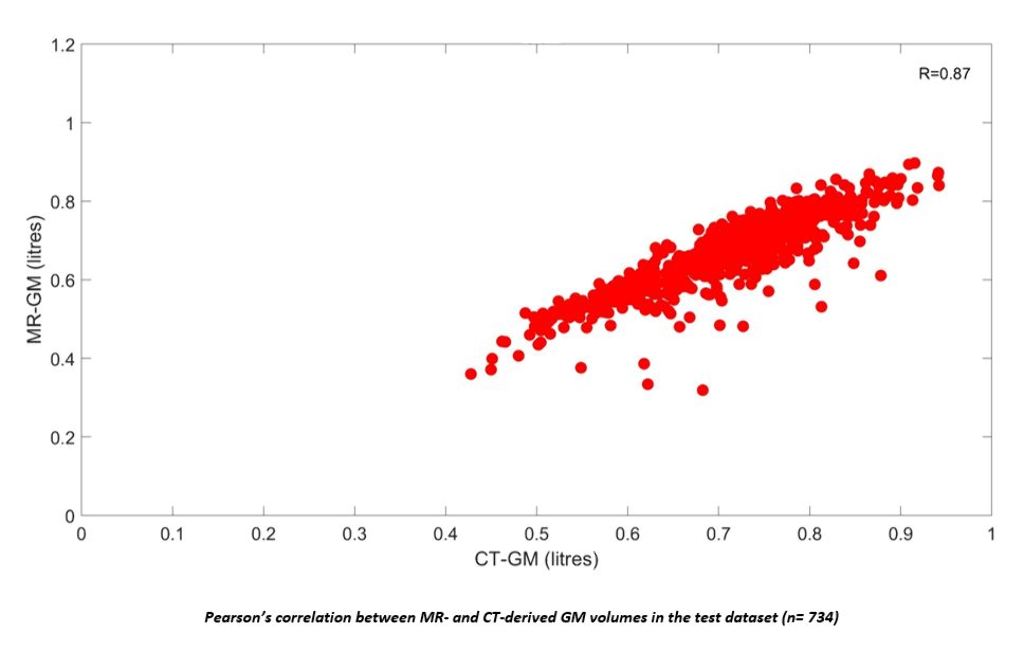
A first dataset was included from the Gothenburg H70 Cohort of which 734 participants (age= 70.42±2.6 years, 52.6% female) had paired CT and T1-weighted MRI scans. Grey matter (GM), white matter (WM), cerebrospinal fluid (CSF) and intracranial volume (ICV) segmentations were derived from MRI. A U-Net was trained on these to automatically predict the same tissue classes from CT, which were compared with MRI-segmentations for volumetric, spatial and shape similarity. Associations between CT-derived GM volumes (controlled for ICV), plasma NfL and mini mental state examination (MMSE) were tested using linear regressions. A second dataset including 300 participants with paired CT and MRI of which 30% had a clinical dementia diagnosis was included from the NUS MACC to validate diagnostic performance of the acquired measures.
Results
In the H70 Cohort, high volumetric correlations and continuous Dice scores were observed between CT- and MRI-derived GM, WM, CSF and ICV. Lower CT-derived GM volumes were significantly correlated with higher plasma NfL (p=0.005) and lower MMSE scores (p= 0.001).


Conclusions
Preliminary results demonstrate clear potential for automated CT-derived volumetry in the assessment of neurodegeneration. Future work will improve the applied methodology, explore associations with visual assessments of GM atrophy, and the concrete diagnostic potential of both CT-volumetry and plasma NfL.
A DEEP-LEARNING BASED FRAMEWORK FOR BRAIN-ATROPHY MEASUREMENT
Abstract
Aims
Image analysis methods based on co-registration of two serial MRI scans, such as the boundary shift integral (BSI) and tensor-based morphometry (TBM), provide precise measurement of volumetric change. However, BSI is semi-automated and restricted to regions with well-defined boundaries, whereas available TBM implementations, such as Advanced Normalization Tools (ANTs), are computationally intensive and slow.
Advancements in artificial intelligence (AI) for deformable image registration allow faster computation without loss of sensitivity. Here, we present an AI-based non-linear registration algorithm to measure longitudinal brain atrophy.
Methods
Our algorithm uses a convolutional neural network and a spatial transformation network and does not require manual labelling for training. It warps one image to another by minimizing their difference, while preserving topology. Voxel-wise volume change is measured using the Jacobian Determinant of the warp field and summed within a specified region of interest. We evaluated our algorithm in ADNI1 participants (24 HC, 23 AD) and assessed the model’s ability to measure 1-year change in whole brain and ventricles. Group comparisons were performed using a Mann-Whitney U-test.
Results
Our algorithm’s registration performance was comparable to ANTs (normalized cross-correlation was 0.983 (SD=0.0065) and 0.9731 (SD=0.017) respectively). Both BSI and our algorithm showed significant group difference for whole brain and ventricles (all p < 0.003), whereas in ANTs there was a significant group effect for ventricles (p<0.001), but not whole brain (p=0.428).
Conclusions
Our algorithm produced fast and accurate non-linear registrations. Our preliminary results also show that the measured atrophy rates were sensitive to clinical diagnosis, comparable to current standard approaches.
RESTING-STATE CO-ACTIVATION PATTERNS AS ACCURATE PREDICTORS OF LATE-STAGE ALZHEIMER’S DISEASE IN MOUSE MODELS.
Abstract
Aims
Alzheimer’s disease (AD), a neurodegenerative disorder marked by accumulation of extracellular amyloid-beta (Aβ) plaques leads to progressive loss of memory and cognitive function. Resting-state fMRI (RS-fMRI) studies have provided links between these observations in terms of disruption of default mode and task-positive resting-state networks (RSNs). The objective of our study was to investigate dynamic fluctuations in RS-fMRI signals in a mouse model (TG2576) of amyloidosis at a late stage (18-months) using sets of resting-state co-activation patterns (CAPs) & to assess their predictive ability.
Methods
CAPs are transient patterns of co-(de)activation that form the basis of RSNs. Here, we followed the approach by Gutierrez-Barragan et al. (2019) that divided all time frames within a scan into spatially similar clusters to extract CAPs from RS-fMRI in 10 TG2576 female mice and 8 wild-type (WT) littermates. Subsequently, we matched the CAPs from two groups using the Hungarian method & compared their temporal (duration, occurrence rate) and the spatial (lateralization of significantly activated voxels) properties. Finally, we tested the predictive ability of CAPs by using their spatial & temporal components as features for training a classifier in order to distinguish the transgenic mice from the WT.
Results
We found robust differences in the spatial component of matched CAPs. We found that while their duration & occurrence rates classified significantly better than the chance-level, BOLD intensities of significantly activated voxels of CAPs turned out to be a significantly better predictive feature achieving a 90% accuracy.
Conclusions
We conclude that RS-CAPs can serve as diagnostic and, potentially, prognostic biomarkers of Alzheimer's disease.


