The Conference will officially run on Central European Time (CET).
To convert to your local time click here.
The viewing of sessions cannot be accessed from this conference calendar.
All sessions are accessible via the Main Lobby on the Virtual Platform.
Thu, 01.01.1970
IRRADIATION OF LUMINESCENCE DOSIMETERS IN STRAY RADIATION FIELD IN LASER-DRIVEN ACCELERATORS
Abstract
Background and Aims
The Extreme Light Infrastructure (ELI) Beamlines is a state-of-the-art laser driven accelerator facility located in the Czech Republic. It will operate the most intense non-military laser system with ultra-high power up to 10 PW, concentrated plasma intensities of up to 1024 Wcm-2, and ultra-short laser pulses of the order of few femtoseconds. Lasers of a several hundred TWs are already capable of producing a large amount of ionizing radiation when interacting with suitable targets. Thus prompts the need for accurate dosimetry in ultra-short pulsed fields; this is a need shared with FLASH radiotherapy facilities alike. Solid-state detectors, such as Thermoluminescence (TL) and Optically Stimulated Luminescence (OSL) detectors are promising technique used for this purpose. They are immune to electromagnetic pulses and the liberated electron-hole pairs are trapped on a 10-15 s to 10-13 s timescale.
Methods
Within the compass of the 18HLT04 UHDpulse EMPIR project, TL and OSL detectors are irradiated in stray radiation fields at ELI Beamlines. Several luminescent materials are used. Exploiting different laser and experimental setups and different distances from the source, it is possible to investigate a wide range of dose rates.
Results
Detector responses are evaluated and compared to Monte Carlo expectations. Preliminary results are presented.
Conclusions
The results show how the irradiated luminescence dosimeters respond in complex radiation fields.
This project 18HLT04 UHDpulse has received funding from the EMPIR programme co-financed by the Participating States and from the European Union's Horizon 2020 research and innovation programme.
PLASTIC SCINTILLATOR UNDER ULTRA-HIGH DOSE RATE ELECTRON BEAM: LONG TERM DAMAGES AND CHANGES IN OPTICAL RESPONSE
Abstract
Background and Aims
Background and aims: Scintillation detectors have advantages that could benefit dosimetry for FLASH radiotherapy, but radiation damages could alter their properties. This study aims at determining long term effects of ultra-high dose rate electron radiation on scintillation dosimeters.
Methods
Methods: Samples of clear and scintillating fibers (1 mm diameter, 15 mm long) were irradiated under electron beams with ultra-high dose per pulse (1 Gy/pulse, 20 MeV, 5 Hz, 2.5 µs pulse length) up to a total dose of 20 kGy. Scintillation and transmission of irradiated samples were assessed using the Hyperscint RP200 platform (Medscint inc., Canada), around 3 months after irradiation.
Results
Results: Emission spectrum remained stable for all doses (figure 1), with a maximum RMS difference of 0.002 between irradiated and unirradiated samples. The total light production and transmission seems minimally affected from irradiation, up to 20 kGy (figure 2). The error bars result from the differences due to sample manufacturing. However, further investigations will have to be made to confirm this statement.
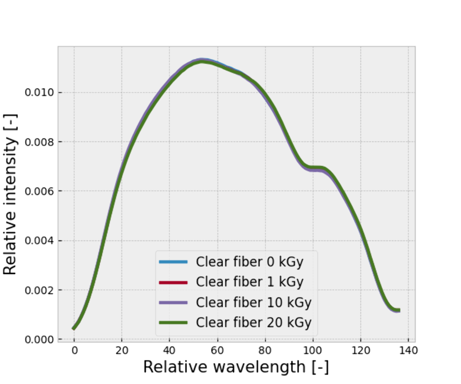
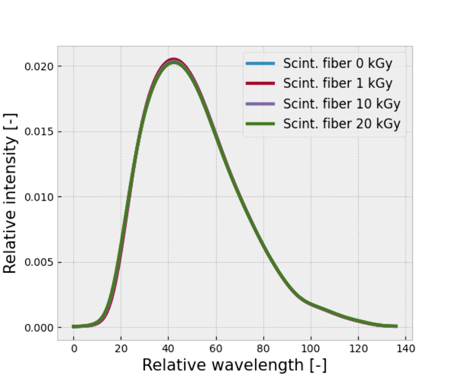
Figure 1: Emission spectra of the irradiated samples.
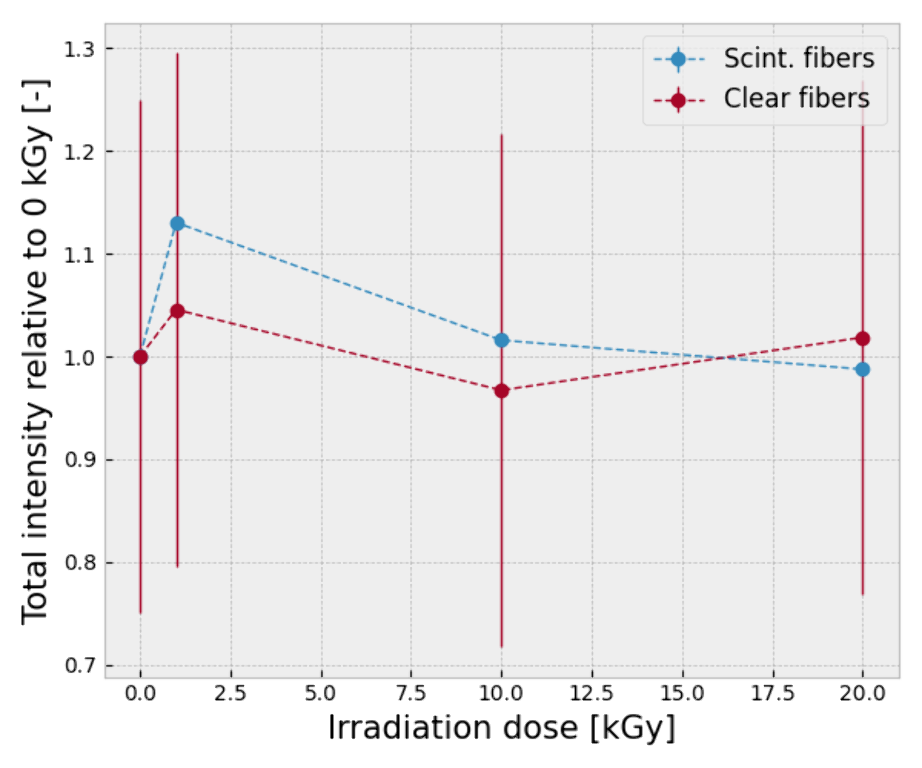
Figure 2: Total intensity measured as a function of total dose.
Conclusions
Conclusions: Scintillating and clear plastic fibers do not show significant long term alterations when irradiated up to 20 kGy with ultra-high dose rate electron beams.
Acknowledgements: This project 18HLT04 UHDpulse has received funding from the EMPIR programme co-financed by the Participating States and from the European Union’s Horizon 2020 research and innovation programme. Special thanks to the team of Medscint inc. for their help in analysing the irradiated samples.
VENTED IONIZATION CHAMBERS FOR ULTRA-HIGH DOSE PER PULS CONDITIONS
Abstract
Background and Aims
Vented ionization chambers (IC) are used as the standard dosimeter for clinical reference dosimetry. For ultra-high dose per pulse (DPP) in the range of 0.6 – 10 Gy, as under investigation for FLASH radiotherapy with electrons, available ICs show large deviations due to ion recombination. However, it is desirable to use ICs also under ultra-high DPP conditions as a secondary standard.
Methods
Parallel plate prototype ICs with different electrode distances d manufactured by PTW were investigated at PTB's research electron accelerator (20 MeV, 5 Hz, 2.5 µs pulse duration). To determine the DPP reference, the beam current monitor was calibrated against alanine. The measurements were compared to a numerical approach by solving a system of partial difference equations, taking into account charge creations by the radiation, their transport and reaction in an applied electric field.
Results
As the electrode distance decreases, the deviations due to ion recombination become smaller. For the prototype with d=0.25mm, almost no deviation is detectable anymore. There is a good agreement between measurements and simulations.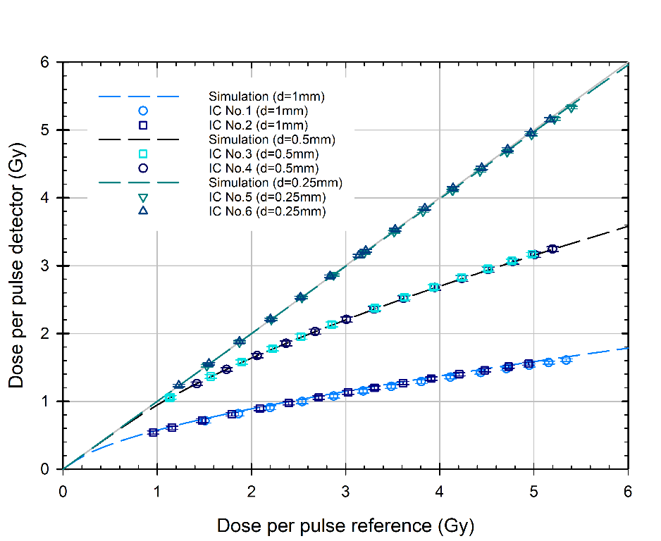
Figure 1: Measured and simulated DPP vs. reference DPP for ICs with different electrode distances d and a voltage of 250V.
Conclusions
Our investigation confirms the previous findings of Gomez et al. presented on this conference. Parallel plate ICs with very small electrode distances are a promising tool for real time dosimetry in FLASH radiotherapy.
Acknowledgement
This project 18HLT04 UHDpulse has received funding from the EMPIR programme co-financed by the Participating States and from the European Union’s Horizon 2020 research and innovation programme.
MULTI-LEAF FARADAY CUP FOR QUALITY ASSURANCE IN RADIATION THERAPY WITH ELECTRON AND ION BEAMS AT CONVETIONAL AND ULTRA-HIGH DOSE RATE
Abstract
Background and Aims
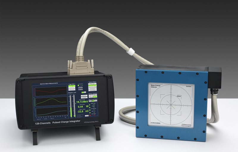
For quality assurance in radiation therapy with high-energy electrons, protons or carbon ions, the beam energy has to be checked in clinical routine. This test is time-consuming and requires expensive equipment. If the dose rate is varied by orders of magnitude to compare ultra-high dose rate with conventional irradiation, one should be able to measure the energy quick and reliable and independently of the accelerator readouts or manufacturer's specifications. For this purpose, a portable Multi-Leaf Faraday Cup (MLFC) was developed (Fig.1), with a readout electronics whose is not influenced by the dose rate.
Methods
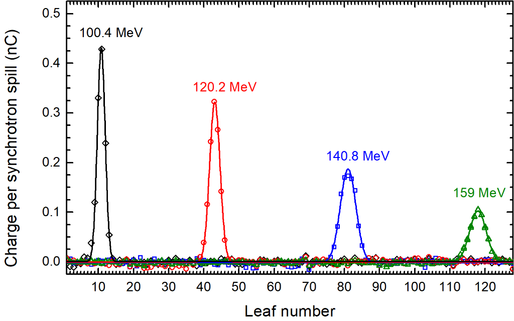
The MLFC was irradiated with protons with energies between 64 – 252 MeV from a synchrotron source. Aluminium disks were placed in front of the MLFC as range shifter. Charge and energy were measured per synchrotron spill (Fig.2). Furthermore, the MLFC was tested in electron beams of conventional and ultra-high dose per pulse.
Results
For monoenergetic electron or proton beams energy differences of 80 keV (0.8%) or 60 keV (0.1%), respectively, can be clearly distinguished.
Conclusions
Due to the measurement principle, which is independent of the dose rate, the MLFC is suitable for quality assurance of charged particle radiation with conventional as well as ultra-high dose rate, for short pulses as well as for continuous radiation. Energy and intensity can be measured quick and reliable.
Acknowledgement: This project 18HLT04 UHDpulse has received funding from the EMPIR programme co-financed by the Participating States and from the European Union’s Horizon 2020 research and innovation programme.
PROOF OF PRINCIPLE OF A PYROELECTRIC CALORIMETER WITH ELECTROMETER READOUT FOR THE ABSOLUTE DOSIMETRY OF FLASH RADIATION
Abstract
Background and Aims
Given the extremely high doses-per-pulse in FLASH radiotherapy, calorimetry-based dosimetry is a proposed alternative to ion chambers, which perform poorly under such conditions. However, to-date, calorimeters require instrumentation for readout that is too complex and specialized for routine clinical use. This work describes the development and initial characterization of a pyroelectric calorimeter capable of FLASH dosimetry measurements that can be read out with only an electrometer.
Methods
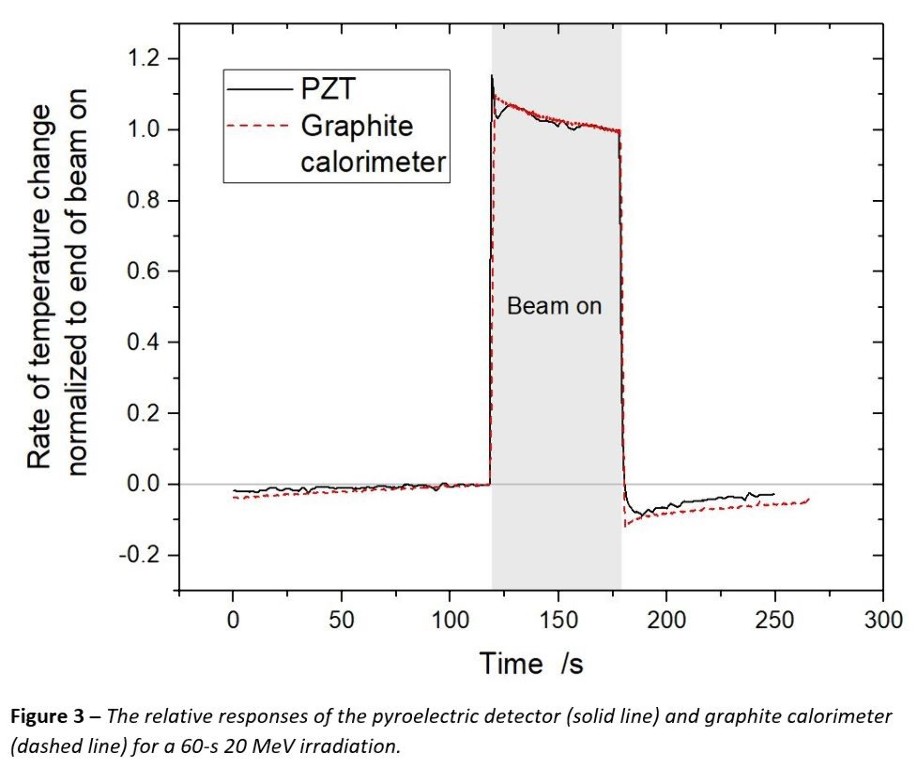
The pyroelectric crystal (lead‑zirconate-titanate; PZT) is a current source with a signal magnitude that is directly proportional to a change in temperature, hence a calorimeter. As an initial characterization, a thermistor was affixed to the crystal and the assembly was embedded in a copper block. The temperature of the copper was then modulated by exposing it to the heat of a halogen lamp, simulating the effect of a FLASH beam. Concurrent pyroelectric and graphite calorimetry measurements were then carried out in a 20 MeV electron beam.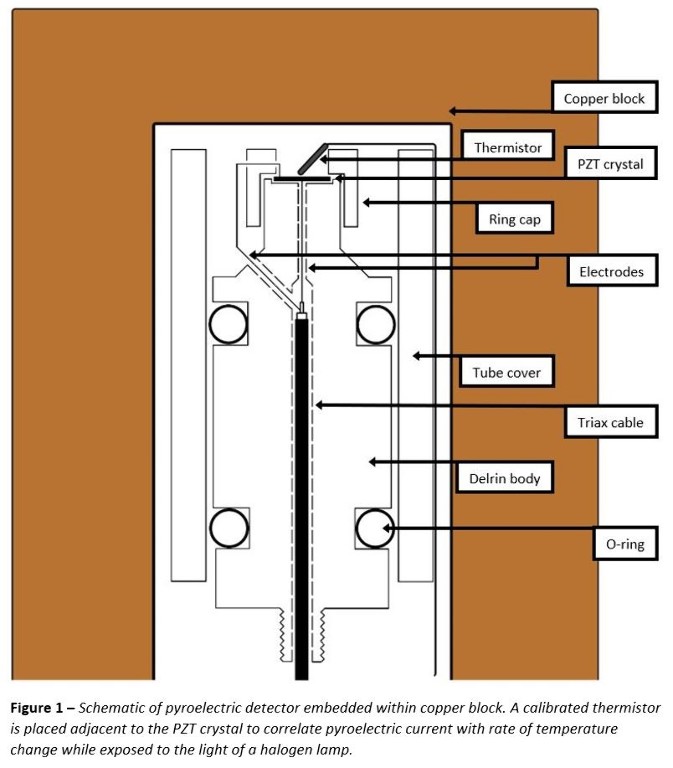
Results
Thermal calibration data spanning rates of temperature change from ± 8 mK∙s-1, or up to 3.5 Gy∙s-1 equivalent dose rate demonstrates that pyroelectric current varies linearly with the time derivative of the measured temperature. When irradiated, the ratio of pyroelectric current to graphite calorimeter dose was consistent to within an RMS of 0.6 %.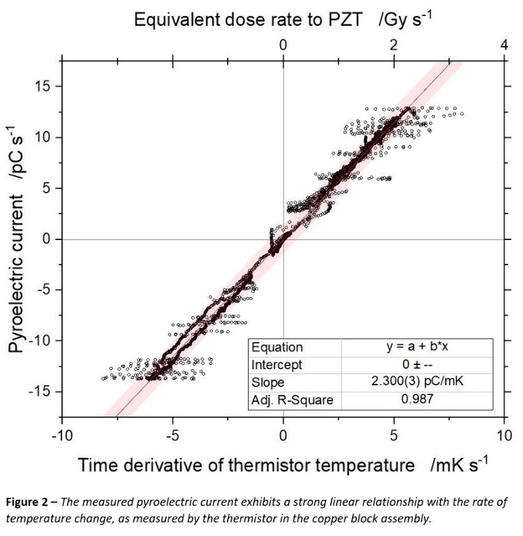
Conclusions
This proof of principle demonstrates the feasibility of operating a pyroelectric calorimeter with an electrometer for the purpose of absolute determination of absorbed dose rates greater than a few Gy∙s‑1, relevant for FLASH dosimetry.
DEVELOPMENTS IN ACTIVE NEUTRON SPECTROMETRY FOR NEUTRON STRAY RADIATION FIELD CHARACTERIZATION IN FLASH RADIOTHERAPY
Abstract
Background and Aims
During FLASH radiotherapy, secondary pulsed radiation fields are created outside of the primary beam. These fields, composed of radiation of different types and/or energy, can cause secondary doses to healthy tissue and critical organs outside of the target volumes. The main component is neutron radiation, contributing significantly to the secondary equivalent dose. For personalized dose management, especially for paediatric patients, the unwanted secondary dose must be reliably estimated and minimized. As a basis for reliable and traceable neutron dosimetry, neutron spectrometry for pulsed fields plays a key role in the proper characterization of the stray radiation fields.
Methods
In the framework of the project UHDpulse, two independent neutron spectrometric systems are being developed, based on Bonner sphere spectrometers (BSS). The first approach consists of adapting the front-end electronics of the LUPIN [M. Caresana et al., NIM A 712 (2013) 15], developed specifically for pulsed neutron fields, for use with a BSS. The second approach builds upon the BSS NEMUS [B. Wiegel and A.V. Alevra, NIM A 476 (2002) 36] with the standard 3He-filled proportional neutron counter replaced by a 235U-coated ionization chamber. Both approaches rely on active detection methods, being able to study in detail the time variation of the pulsed neutron fields.
Results
This contribution discusses both approaches and summarizes experimental tests.
Conclusions
An outline of future developments is presented.
This project 18HLT04 UHDpulse has received funding from the EMPIR programme co-financed by the Participating States and from the European Union’s Horizon 2020 research and innovation programme.

