Presenter of 1 Presentation
DEEP LEARNING BASED LVO DETECTION ON CT ANGIOGRAPHY OF BRAIN
Abstract
Background and Aims
The early detection of anterior circulation large vessel occlusion (LVO) by computed tomography angiogram (CTA) is critical for treating acute ischemic stroke with mechanical thrombectomy.
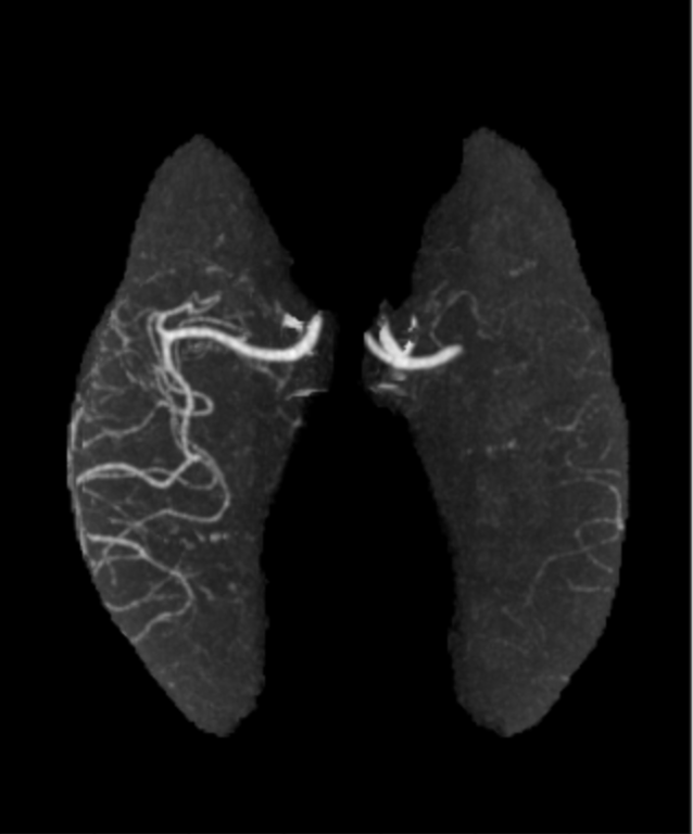
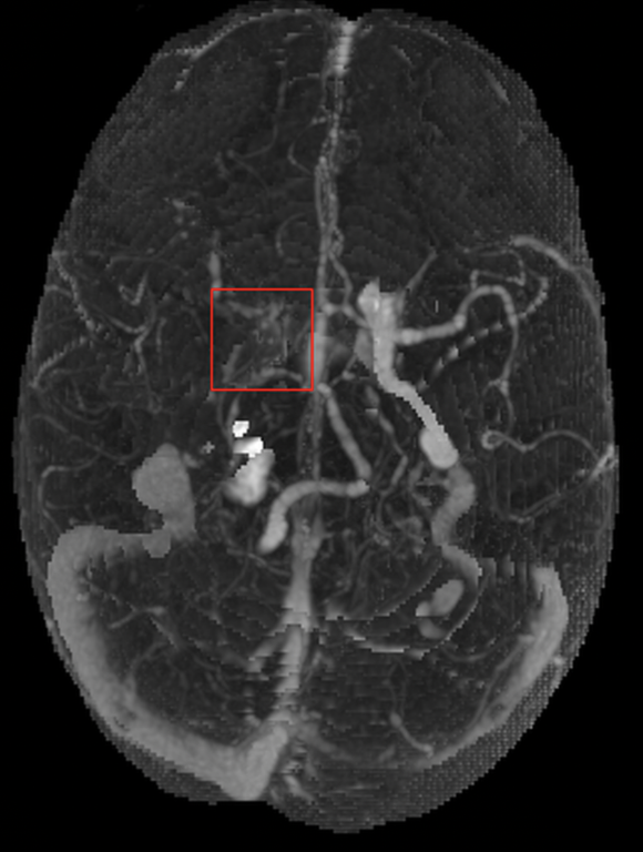
We present a deep learning solution that detects Internal Carotid Artery (ICA) and M1/M2 Middle Cerebral Artery (MCA) occlusions on the maximum intensity projections (MIPs) of the brain's vascular territories (VTs).
Methods
Each CTA is preprocessed using a classifier which trims off the neck and keep only the brain.
A neurologist annotated the brain's left and right side of MCA and ICA territories on a single CTA. This is used as a reference to annotate VT maps on 942 CTAs.
These CTA scans were used to train and validate a deep learning based segmentation model which outputs brain VT masks and is aligned on corresponding CTA to get VT MIPs.
Deep learning based classification with segmentation models were trained on VT MIPs of 1083 MCA LVO positive scans, 840 ICA LVO positive scans, and 5142 LVO negative scans.
Results
Dice Similarity Coefficient (DSC) was adopted to validate the output of the segmentation model. VT outputs were validated on 232 CTAs with a DSC of 0.90.
| Validation Set | ||
| MCA LVO Detection | No. of positive scans | 445 |
| No. of negative scans | 2155 | |
| Sensitivity | 0.88 | |
| Specificity | 0.91 | |
| ICA LVO Detection | No. of positive scans | 360 |
| No. of negative scans | 2155 | |
| Sensitivity | 0.92 | |
| Specificity | 0.89 |
Conclusions
Even with the contrast contamination of veins, MIPs are not affected, and hence the deep learning model effectively and efficiently detects the ICA and MCA occlusions.


