Author Of 1 Presentation
REAL-TIME OPTICAL OXIMETRY UNDER IRRADIATION
Abstract
Background and Aims
Transient changes in oxygen tension in tissues taking place during FLASH radiotherapy may explain its biological effects. However, because the kinetics of oxygen depletion and recovery occur on a very short timescale, it is challenging to measure these effects in vivo using existing methods. Here we developed a real-time optical oximetry system with millisecond temporal resolution to elucidate early radiochemistry under irradiation.
Methods
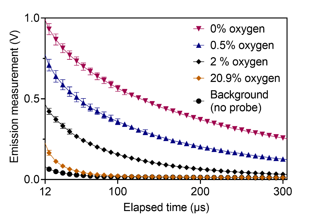
Oxygen measurements were performed in vitro using the phosphorescence quenching method and a water-soluble molecular nanoprobe (Fig. 1). An epifluorescence fiber-coupled system was designed and built. The system was validated using a standard dissolved oxygen meter. The changes in oxygen per unit dose (G-value) were quantified in response to irradiation by 320 kVp x-ray and 16 MeV electron beam at dose rates ranging from 0.04 Gy/s to 100 Gy/s.

Results
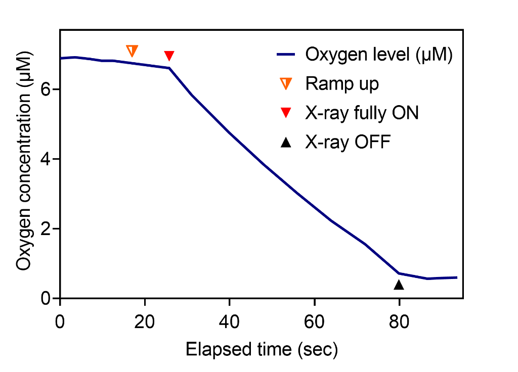
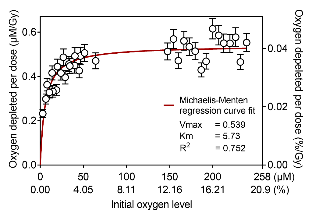
Transient oxygen depletion of phosphate buffer solution under standard kV X-ray irradiation was continuously measured every millisecond (Fig. 2). The samples at normoxia with oxygen concentration of 150–240 µM had constant G-value of 0.54 uM/Gy, however hypoxic samples (15 µM and below) had significantly lower G-values (Fig. 3).


Conclusions
Our observations suggest that oxygen depletion rate decreases under hypoxia, with the measured data being a good fit to Michaelis-Menten kinetics. Future works will measure oxygen depletion kinetics of biomimetic lipid emulsions and in vivo mice under irradiation. We anticipate that the proposed method will capture crucial millisecond-order oxygen change after individual e-beam pulses.

