The Conference will officially run on Central European Time (CET).
To convert to your local time click here.
The viewing of sessions cannot be accessed from this conference calendar.
All sessions are accessible via the Main Lobby on the Virtual Platform.
Thu, 01.01.1970
COMPARISON OF H2O2 AND HO· PRIMARY YIELDS AND O2 DEPELTION AFTER IRRADIATION AT UHDR AND CONVENTIOANL-DOSE RATE WITH 6MEV ERT6/ORIATRON
Abstract
Background and Aims
Ultra-high dose rate (UHDR) irradiation produces the FLASH effect (anti-tumor effect without normal tissue toxicity) at average dose rates above 100 Gy/s. Two physico-chemical scenarios were proposed as a mechanistic basis for the FLASH effect following water radiolysis: 1. Altered radical-radical reactions and 2. Depletion of O2 by free radicals. To investigate these questions, we determined primary radiolytic yields (G°-values) of hydrogen peroxide and hydroxyl radicals. G(H2O2) was also determined in low O2 condition (1%), intermediate levels (4%) and atmospheric conditions (21%) following homogenous phase. O2 depletion was also measured.
Methods
Scavenging methods were used to estimate radiolytic yields of H2O2 in water samples. Hydroxyl radicals production was estimated using EPR spin trapping with DMPO. O2 measurements were performed using OxyLite probe.
Results
When UHDR and CONV-irradiation were compared, similar primary yields of H2O2 were found and EPR measurements suggested no differences in HO· production. However, G(H2O2) was significantly lower after irradiation at UHDR in samples equilibrated at 4%. O2 measurements resulted in similar but low depletion with both modalities at intermediate and atmospheric O2 conditions, whereas at low O2 level, oxygen depletion was lower at UHDR.
Conclusions
These observations suggest that initial radiation chemistry is similar in both modalities: Similar yields of radicals ‘‘escaping’ ’track recombination after ionization. However, irradiation at UHDR produces less H2O2 at intermediate O2 levels following initial chemistry events, supporting occurrence of scenario 1. O2 depletion hypothesis is not favored by results obtained in pure water.
Acknowledgement: The study is funded by FNS Synergia grant (FNS CRS II5_186369)
OXYGEN DEPLETION IN ULTRA-HIGH DOSE RATES FOR PROTONS AND ELECTRONS: EXPERIMENTAL APPROACH IN WATER AND BIOLOGICAL SAMPLES
Abstract
Background and Aims
In FLASH radiotherapy (RT), a protective effect of healthy tissue was observed, while tumor control remains comparable to conventional RT. One possible explanation is the oxygen depletion hypothesis, in which radiolysis of water/cytoplasma causes the production of radicals that then react with O2 dissolved in water. This would cause a reduction in O2, which results in a hypoxic target and thus a radio-protective effect. In a previous study (Jansen et al. 2021), we measured O2 depletion using an optical sensor in sealed water phantoms during irradiation at high dose rates (<300 Gy/s) for protons, carbon ions and photons.
Methods
In the study presented here, this experiment was conducted further to ultra-high dose rates (10^9 Gy/s) with protons at DRACO and electrons at ELBE, where also the impact of different pulse structures on O2 depletion in water was tested. In addition, various settings were tested in order to irradiate zebrafish embryos with FLASH while simultaneously measuring O2.
Results
We were able to confirm the results of our previous study even at ultra-high dose rates and with electrons. Furthermore, it was possible to measure O2 depletion during zebrafish embryo irradiation making a simultaneous study of biological response and O2 depletion possible.
Conclusions
Not enough O2 was depleted at clinical doses to explain a FLASH effect based on radiation-induced hypoxia. The amount of O2 depleted per dose depends on dose rate, and higher dose rates deplete slightly less O2. The experimental set up allows for future joint experiments of biological samples and oxygen monitoring.
MODEL STUDIES OF THE ROLE OF OXYGEN IN THE FLASH EFFECT
Abstract
Background and Aims
Convincing evidence has been provided that lowering the mean dose rate below 30 Gy/s (Montay-Gruel et al. Radiother Oncol 124: 365-369, 2017) or increasing the oxygen tension through carbogen breathing (Montay-Gruel et al. PNAS 116: 10943-10951, 2019) can reduce (if not eliminate) the sparing effect of FLASH in the mouse brain. Three theoretical models have since been proposed to account for the dependence of FLASH on oxygen, namely, (i) Radiation-induced transient oxygen depletion (TOD), (ii) Cell-specific differences in the ability to detoxify and/or recover from injury caused by reactive oxygen species, (iii) Self-annihilation of radicals by bimolecular recombination.
Methods
These theoretical models were confronted to experimental evidence relating to physical chemistry and the response of biological systems to ultrahigh dose rate irradiation in light of the chemical reactions underlying the radiosensitizing effect of O2 from immediate, early processes to late complications.
Results
The radiosensitizing effect of oxygen has long been known to stem from peroxyl radicals (ROO•). Peroxyl radicals areformed by the addition of O2 in its fundamental, triplet state to short-lived carbon-centered radicals (R•) generated upon H• atom abstraction from aliphatic, unsaturated or conjugated substrates (RH).
Conclusions
Any attempt at deciphering the physical-chemical processes that underpin the FLASH effect must take into consideration the fate of these radicals, and in particular the probability of radical recombination. The radiation dose rate emerges as the most important factor. We propose testable procedures to investigate the mechanisms that underly the FLASH effect in normal cells, and differential tumor vs. normal cells responses.
ULTRAFAST TRACKING OF OXYGEN DYNAMICS DURING PROTON FLASH
Abstract
Background and Aims
Radiotherapy at ultrahigh dose rates (FLASH, >40Gy/sec) compared to conventional radiotherapy (CR; <1 Gy/sec) has been shown to provide an advantage by selective protection of normal versus tumor tissue. Depletion of molecular oxygen causing radioprotection has been proposed to underpin the FLASH effect, however no experimental data have been presented to test this hypothesis. Our goal was to assess the oxygen dynamics during and following proton FLASH versus CR.
Methods
Measurements of oxygen were performed in vitro (solutions in sealed vials) and in vivo in mice (healthy leg muscle and tumors) using the phosphorescence quenching method with probe Oxyphor PtG4 at rates reaching 3 measurements/millisecond during proton irradiation delivered with CR or FLASH dose rates.
Results
In closed systems in vitro the rate of oxygen depletion is oxygen-independent for both FLASH and CR when [O2]>~70 μM. The g-values are lower for FLASH than for CR by ~15%. In vivo oxygen is depleted upon FLASH with rates that correlate with baseline pO2 (partial pressure of oxygen) values throughout the entire pO2 range: at 30-40 mmHg (normal tissue) a 30 Gy FLASH (100 Gy/s) results in ΔpO2 of -8 mmHg, while at 5-7 mmHg (tumor) ΔpO2 is only -1 mmHg.
Conclusions
This study features the first demonstration of the phosphorescence quenching oximetry at ultrafast rates and in the presence of proton radiation. Oxygen depletion by FLASH in vitro was found to be smaller than by CR, and appears to correlate with baseline pO2 values, being greater for normal tissue than for tumors.
UHDR PROTON BEAM VS. CONVENTIONAL: HYDROGEN PEROXIDE AS FLASH EFFECT SENSOR
Abstract
Background and Aims
Several models assume that delivering radiation at ultra-high dose rates (UHDR) reduces the yields of reactive oxygen species (ROS). Montay-Gruel et al [1] measured a significant decrease of Hydrogen Peroxide (H2O2) after UHDR irradiation compared to conventional dose-rate irradiation (CONV) with electrons. This study aims to verify this assumption in proton beams. We study the UHDR radiation chemistry by the measurement of H2O2 produced by the water radiolysis. Fricke dosimeter is used as dose control and as ROS sensor as well.
Methods
Using ARRONAX facility, we produced proton beams (68MeV) ranging from low (0.25Gy/s, 100Hz, pulse dose rate=6.3Gy/s) to ultra-high dose rates (7500Gy/s, single pulse). Doses ranging from 5Gy to 80Gy were delivered to 1.5ml Eppendorf tubes filled with water. In this investigation, Fricke dosimetry [2] is used to verify the dose for both irradiation modes. The second step is the determination of H2O2 concentrations after irradiation with the Ghormley triiodide method [3].
Results
In this work, we have brought the evidence of the Flash effect by the H2O2 measurement in the two cases: (i) For CONV irradiations, we determined a H2O2 chemical yield close to the literature at 0.9 10-7 mol.J-1 [4], (ii) For UHDR irradiations, this yield is measured to a significant lower value. Observed Fricke values are the same for both modes.
Conclusions
We have revealed a radio-chemical FLASH effect for the proton beam by the H2O2 measurement as radiolytic species during the irradiation of water. Further studies will be performed to determine the beam time structure effect.
PLASMID DNA DAMAGES AFTER FLASH VS CONVENTIONAL DOSE RATE IRRADIATIONS IN VARIOUS OXYGEN CONDITIONS
Abstract
Background and Aims
In our work we thought to compare the effects of conventional (CONV) vs ultra-high dose rate (UHDR) by quantifying DNA strand breaks (SB) after irradiation of plasmid-DNA (pBR322) under various oxygen concentrations.
Methods
Supercoiled pBR322 was irradiated dry or in water using a 6 MeV FLASH-validated electrons beam, with increasing doses (1-100 Gy) and dose per pulse (0.01 Gy/s (CONV), 5.0*102 to 5.6*106 Gy/s (UHDR)) and at atmospheric (21%), physoxic (4%) and hypoxic (0.5%) oxygen level. The increase of relaxed (R) and linear (L) plasmid forms after irradiation was quantified by agarose gel electrophoresis and used to compute single and double SB yields.
Results
Dry, atmospheric conditions cause similar yields of SB in CONV and UHDR. Aqueous conditions shows higher SB yields as expected. Physoxia induces radioprotection compare to atmospheric condition: 50% of R at 10 Gy (4% O2) vs 2 Gy (21% O2), but no difference relative to dose rate. Hypoxia revealed higher SB yields than physoxia in CONV (50% of R at 6 Gy) but 2x less SB in UHDR for doses >30 Gy (see figure for L).

Conclusions
First results in dry condition suggest that direct effects are not involved in FLASH. In aqueous condition, 4% oxygen mimicking healthy tissues shows no difference between UHDR and CONV, while 0.5% oxygen mimicking tumors shows less damages in UHDR. These results are opposite to the preclinical results showing the FLASH effect. Thus, plasmid irradiation might be useful to understand DNA damage at UHDR but seems barely relevant to investigate the FLASH effect at the biological level.
REAL-TIME OPTICAL OXIMETRY UNDER IRRADIATION
Abstract
Background and Aims
Transient changes in oxygen tension in tissues taking place during FLASH radiotherapy may explain its biological effects. However, because the kinetics of oxygen depletion and recovery occur on a very short timescale, it is challenging to measure these effects in vivo using existing methods. Here we developed a real-time optical oximetry system with millisecond temporal resolution to elucidate early radiochemistry under irradiation.
Methods
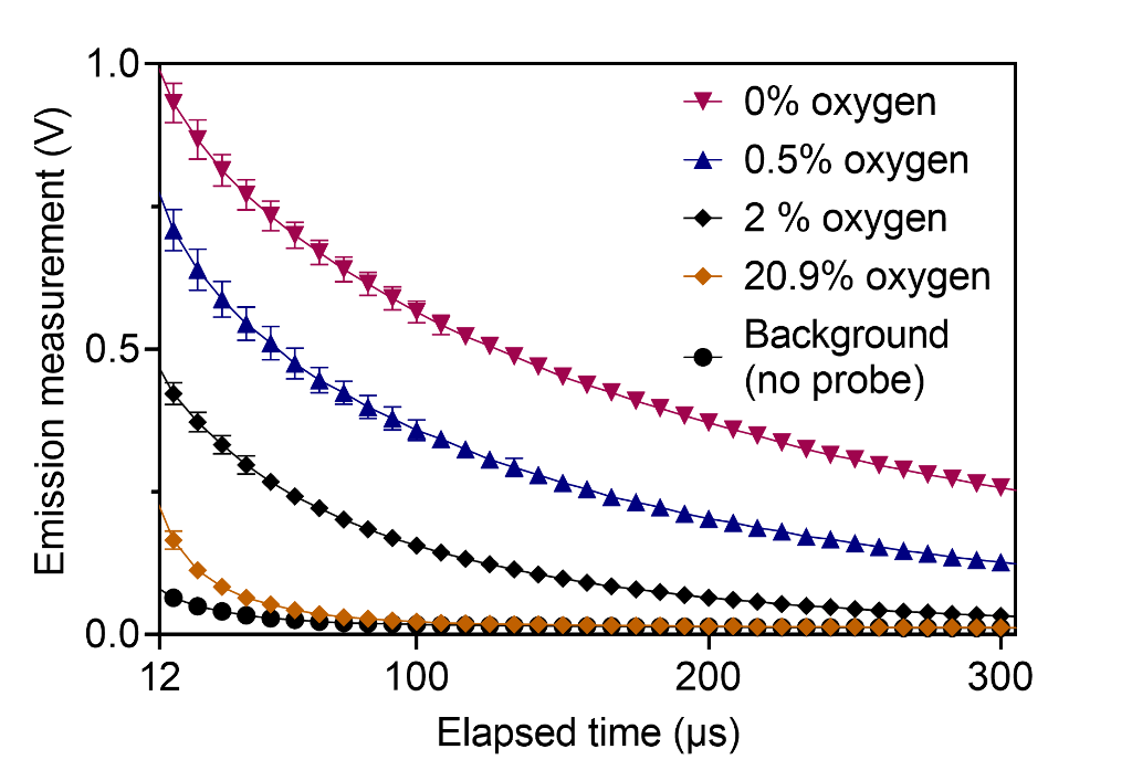
Oxygen measurements were performed in vitro using the phosphorescence quenching method and a water-soluble molecular nanoprobe (Fig. 1). An epifluorescence fiber-coupled system was designed and built. The system was validated using a standard dissolved oxygen meter. The changes in oxygen per unit dose (G-value) were quantified in response to irradiation by 320 kVp x-ray and 16 MeV electron beam at dose rates ranging from 0.04 Gy/s to 100 Gy/s.

Results
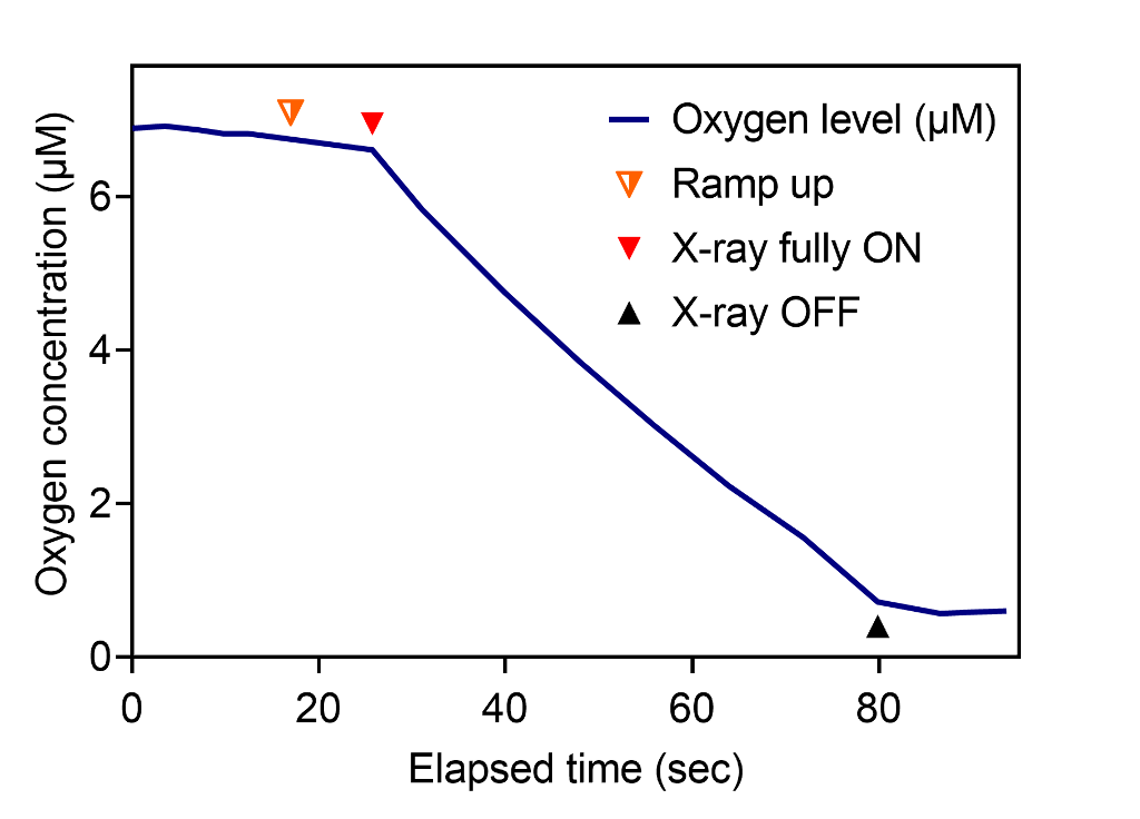
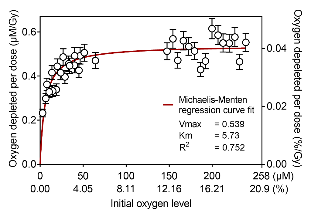
Transient oxygen depletion of phosphate buffer solution under standard kV X-ray irradiation was continuously measured every millisecond (Fig. 2). The samples at normoxia with oxygen concentration of 150–240 µM had constant G-value of 0.54 uM/Gy, however hypoxic samples (15 µM and below) had significantly lower G-values (Fig. 3).


Conclusions
Our observations suggest that oxygen depletion rate decreases under hypoxia, with the measured data being a good fit to Michaelis-Menten kinetics. Future works will measure oxygen depletion kinetics of biomimetic lipid emulsions and in vivo mice under irradiation. We anticipate that the proposed method will capture crucial millisecond-order oxygen change after individual e-beam pulses.

