Lars-Olof Wahlund, Sweden
Karolinska Institutet Department of Neurobiology, Care Sciences, and SocietyAuthor Of 1 Presentation
FIRST-LEVEL ASSESSMENT OF NEURODEGENERATION USING DEEP-LEARNING BASED BRAIN CT SEGMENTATIONS AND BLOOD-DERIVED MEASURES OF NEUROFILAMENT LIGHT
Abstract
Aims
Brain computed tomography (CT) and blood sampling are two routinely performed examinations in the clinical assessment of neurodegenerative diseases. We aim to evaluate the diagnostic performance of automated CT-based volumetry, derived with a novel deep-learning approach, and plasma-based neurofilament light chain (NfL).
Methods
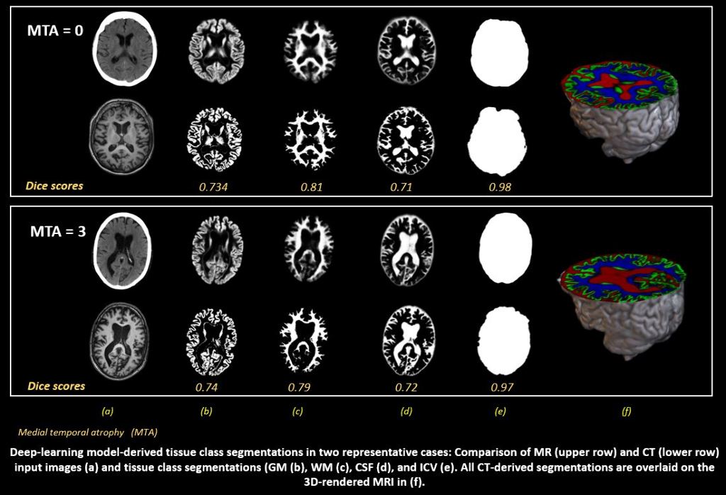
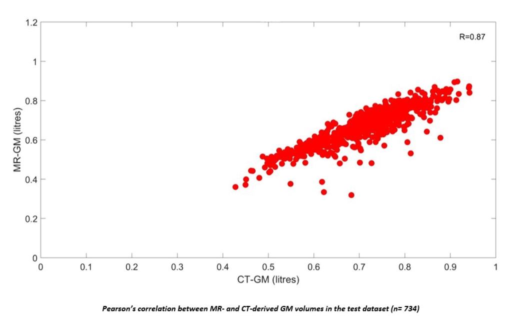
A first dataset was included from the Gothenburg H70 Cohort of which 734 participants (age= 70.42±2.6 years, 52.6% female) had paired CT and T1-weighted MRI scans. Grey matter (GM), white matter (WM), cerebrospinal fluid (CSF) and intracranial volume (ICV) segmentations were derived from MRI. A U-Net was trained on these to automatically predict the same tissue classes from CT, which were compared with MRI-segmentations for volumetric, spatial and shape similarity. Associations between CT-derived GM volumes (controlled for ICV), plasma NfL and mini mental state examination (MMSE) were tested using linear regressions. A second dataset including 300 participants with paired CT and MRI of which 30% had a clinical dementia diagnosis was included from the NUS MACC to validate diagnostic performance of the acquired measures.
Results
In the H70 Cohort, high volumetric correlations and continuous Dice scores were observed between CT- and MRI-derived GM, WM, CSF and ICV. Lower CT-derived GM volumes were significantly correlated with higher plasma NfL (p=0.005) and lower MMSE scores (p= 0.001).


Conclusions
Preliminary results demonstrate clear potential for automated CT-derived volumetry in the assessment of neurodegeneration. Future work will improve the applied methodology, explore associations with visual assessments of GM atrophy, and the concrete diagnostic potential of both CT-volumetry and plasma NfL.


