Presenter of 1 Presentation
INDIVIDUAL PULSE MONITORING AND FEEDBACK SYSTEM FOR FLASH-RT BEAM CONTROL USING FIBER-COUPLED SCINTILLATING DETECTORS
Abstract
Background and Aims
FLASH sources lack dose rate independent dosimeters and dose feedback systems. We developed an ultra-high dose rate beam monitoring system for FLASH-RT, including dose-rate independent scintillating detector and fast electronics.
Methods
A commercially available plastic scintillator and a liquid fluorescein solution were coupled to a gated integrating amplifier and a field programmable gate array (FPGA) for dose monitoring and feedback control. The FPGA was programmed to integrate dose and measure pulse width for each radiation pulse. The detectors were characterized in terms of the radiation stability, mean dose-rate (Ḋm), and dose per pulse (Dp) linearity.
Results
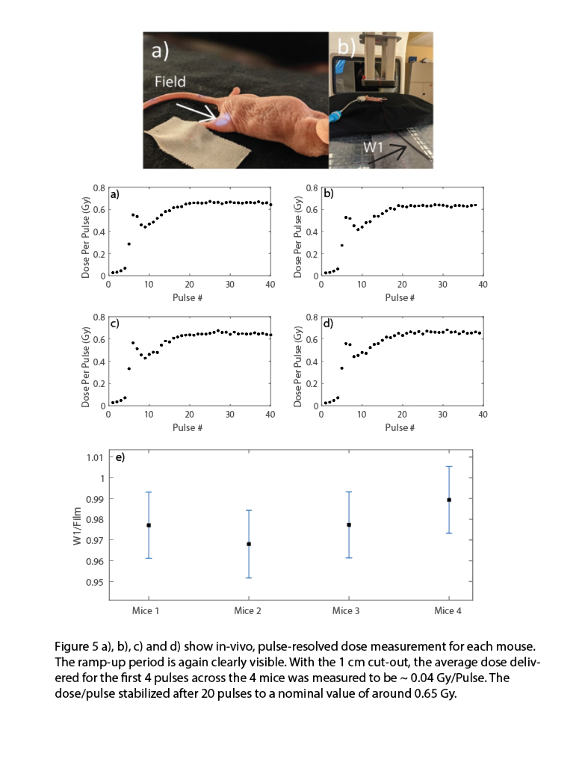
The Dp and the pulse width showed a consistent ramp-up period of ~4-5 pulse. The plastic scintillator was shown to be linear with Ḋm (40-380 Gy/s) and Dp (0.3-1.3 Gy/Pulse) to within ± 3%. However, the plastic scintillator was subject to significant radiation damage for the first 2 kGy (24%/kGy). The fluorescein solution was also tested to be linear with Ḋm (± 3%) and exhibited minimal radiation damage for an initial cumulative dose of 400 Gy. In-vivo dosimetry with a 1 cm circular cut-out revealed that the for the linac ramp-up period of 4 pulses, the average Dp was 0.043 ± 0.002 Gy/pulse, whereas after the ramp-up it stabilized at 0.65 ± 0.01 Gy/Pulse.


Conclusions
The tools presented in this study can be used to determine the temporal beam parameter space pertinent to the FLASH effect. Additionally, the hardware can be used to provide real-time feedback to the linac in terms of direct measurement of dose.
Author Of 3 Presentations
INDIVIDUAL PULSE MONITORING AND FEEDBACK SYSTEM FOR FLASH-RT BEAM CONTROL USING FIBER-COUPLED SCINTILLATING DETECTORS
Abstract
Background and Aims
FLASH sources lack dose rate independent dosimeters and dose feedback systems. We developed an ultra-high dose rate beam monitoring system for FLASH-RT, including dose-rate independent scintillating detector and fast electronics.
Methods
A commercially available plastic scintillator and a liquid fluorescein solution were coupled to a gated integrating amplifier and a field programmable gate array (FPGA) for dose monitoring and feedback control. The FPGA was programmed to integrate dose and measure pulse width for each radiation pulse. The detectors were characterized in terms of the radiation stability, mean dose-rate (Ḋm), and dose per pulse (Dp) linearity.
Results
The Dp and the pulse width showed a consistent ramp-up period of ~4-5 pulse. The plastic scintillator was shown to be linear with Ḋm (40-380 Gy/s) and Dp (0.3-1.3 Gy/Pulse) to within ± 3%. However, the plastic scintillator was subject to significant radiation damage for the first 2 kGy (24%/kGy). The fluorescein solution was also tested to be linear with Ḋm (± 3%) and exhibited minimal radiation damage for an initial cumulative dose of 400 Gy. In-vivo dosimetry with a 1 cm circular cut-out revealed that the for the linac ramp-up period of 4 pulses, the average Dp was 0.043 ± 0.002 Gy/pulse, whereas after the ramp-up it stabilized at 0.65 ± 0.01 Gy/Pulse.


Conclusions
The tools presented in this study can be used to determine the temporal beam parameter space pertinent to the FLASH effect. Additionally, the hardware can be used to provide real-time feedback to the linac in terms of direct measurement of dose.
ELECTRON FLASH FOR THE CLINIC: LINAC CONVERSION, COMMISSIONING AND TREATMENT PLANNING
Abstract
Background and Aims
We present the rigorous commissioning of a modified LINAC to deliver ultrahigh dose-rate (UHDR) electron beam and implementation of its model in a widely adopted treatment planning system (TPS) with minimal changes to the clinical setting.
Methods
A Varian Clinac 2100C/D was converted to deliver UHDR beams by withdrawing the target and scattering foil in 10MV x-ray mode. Beam characteristics and stability were quantified by film, Cherenkov, and scintillation imaging. The Geant4 generated beam model was validated with film and implemented in Varian Eclipse TPS. Electron FLASH radiotherapy (eFLASH-RT) plans were generated for representative mammal and human patient cases accounting for complex geometries and anatomical inhomogeneities.
Results
The surface mean-dose-rate at the isocenter was >230Gy/s for all measured fields with adequate long-term stability (deviations of output <7%, symmetry/flatness <2%, spatial shift and FWHM <2mm). The TPS model was validated to clinical accuracy (average error <1.5% for lateral profiles and <2% for percent-depth-dose profiles). Treatments plans were generated and accurately delivered to normal porcine skin surface tissue and a melanoma tumor in a canine’s posterior oral cavity. A human eFLASH-RT plan comparable to a conventional electron plan was achieved by utilizing routine accessories, oblique gantry angle and couch kick.


Conclusions
Treatment planning and accurate delivery of eFLASH-RT were feasible in minimally modified radiation oncology clinical settings. The modifications and open-source TPS model are readily transferable to facilitate clinical translation of eFLASH-RT.
Acknowledgment: This work was supported by the Norris Cotton Cancer Center (grant P30CA023108) and Thayer School of Engineering (seed funding and grant R01EB024498).
IN VIVO QUANTIFICATION OF OXYGEN DEPLETION BY ELECTRON FLASH IRRADIATION
Abstract
Background and Aims
The major hypothesis for the underlying mechanism of normal tissue sparing by FLASH has focused on oxygen depletion, however no experimental data have been presented to support it. The aim of this study was to assess changes in tissue oxygenation in vivo produced by FLASH irradiation.
Methods
Oxygen measurements were performed in vivo and in vitro using the phosphorescence quenching method and molecular probe Oxyphor 2P. The changes in oxygenation were quantified in response to irradiation by a 10 MeV electron beam operating at either ultra-high dose rates (UHDR) reaching 300 Gy/s or at conventional dose rates of 0.1 Gy/s.
Results
In vitro experiments with 5% BSA solutions resulted in oxygen depletion g-values of 0.19-0.21 mmHg/Gy for conventional irradiation and 0.16-0.17 mmHg/Gy for UHDR irradiation. In vivo, the total decrease in oxygen after a single fraction of 20 Gy FLASH irradiation was 2.3±0.3 mmHg in normal tissue and 1.0±0.2 mmHg in tumor tissue (p-value < 0.00001), while no changes in oxygenation were observed from a single fraction of 20 Gy applied at conventional dose rates.
Conclusions
In vitro experiments with 5% BSA solutions resulted in oxygen depletion g-values of 0.19-0.21 mmHg/Gy for conventional irradiation and 0.16-0.17 mmHg/Gy for UHDR irradiation. In vivo, the total decrease in oxygen after a single fraction of 20 Gy FLASH irradiation was 2.3±0.3 mmHg in normal tissue and 1.0±0.2 mmHg in tumor tissue (p-value < 0.00001), while no changes in oxygenation were observed from a single fraction of 20 Gy applied at conventional dose rates.

